Acne Necrotica Miliaris
&
Acne Necrotica Varioliformis
Acne Necrotica – Photomicrograph X 50
© Prof B Stevens
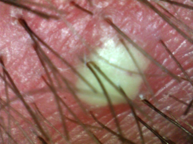
Acne Necrotica
© Prof B Stevens
Variants of this pustular folliculitis affecting the scalp in adults.
I) Acne Necrotica Miliaris – The superficial form of this disease.
Acne Necrotica Miliaris (syn. proprionibacterium) The more superficial form of this disease presents as numerous small vesico-pustules on the scalp often at the central/posterior vertices possibly associated with staphylococcus aureus, demodex folliculorum (tiny mites which live in the hair-follicle) and yeasts of malassez.
Pruritus may co-exist.
The pustules rupture leaving a crust (often the only diagnostic feature).
Patients are often adult males in middle age.
Culture may indicate the presence of Corynebacterium (Propioni-bacterium) or Staphylococcus Aureus. Follicular necrosis usually results.
2) Acne Necrotica Varioliformis – the deeper scarring form of the disease.
Presenting as inflammatory pruritic follicular papules which rupture leaving a crust and scarring.
Treatments:
Topical antibiotics e.g. fusidic acid gel, erythromycin solution. Oral antibiotics e.g. long term tetracycline
Mild topical steroid lotions or creams.
Oral antihistamines
Oral isotretinoin – long term low dose treatment may be tried.
© Prof. B Stevens FTTS
Acne Necrotica Miliaris & Varioliformis
Inga Zemite MD MTTS
Introduction.
Acne necrotica (AN) (folliculitis necrotica) belongs to the group of primary Cicatricial Alopecias. In this working classification diseases are distinguished based on predominant inflammatory cells involved in the process.
2.2. History and Epidemiology.
AN was first described by Bazin in the year 1851. The varioliformis form (described by Hebra) – a condition with the pock-like scars left in the wake of active disease. Sabouraud in 1928 and Lane 1933 described a mild, non-scarring variant of the condition and named it acne necrotica miliaris. Opinions suggest two forms of acne necrotica (acne necrotica varioliformis and acne necrotica miliaris). Some authors believe that acne necrotica or folliculitis necrotica also exists and should not be confused with acne necrotica miliaris as the latter is a non scarring superficial folliculitis that is characterized by minute, very itchy pustules within the scalp while the acne necrotica varioliformis is scarring alopecia form.
AN is a rare condition that usually has its onset in adulthood
2.3. Pathogenesis.
The etiopathogenesis of AN is unknown.
As main factors described or involved in the pathogenesis of disease are
- Propionibacterium acnes and Staphylococcus aureus folliculitis,
- staphylococcus toxin,
- host hypersensitivity to folliculitis and
- rosacea like process.
Theories suggest that the condition maybe an abnormal host response to the bacterium Staphylococcus Aureus or Propionibacterium Acnes folliculitis.
Necrotic excoriation or erosion / destruction of the skin by mechanical insult like a scratching, abrasion or rubbing of the skin prompted by an underlying folliculitis could be another cause of acne necrotica.
2.4. Clinical features.
Mainly adults are affected by AN.
In AN varioliformis, the frontal scalp and adjacent upper forehead are characteristically involved. Other regions of the face and scalp, and the neck and trunk, in a seborrheic distribution, may also be involved. Disease has a chronic, relapsing course.
AN presents in adults as pruritic or painful follicular papules – small, solid, usually inflammatory elevations of the skin that do not contain pus; or follicular papulo-pustules. There are on the skin pruritic or tender, juicy 2-6 mm red to reddish brown follicular papules and papulo pustules that within days umbilicate and undergo central necrosis, develop central hemorrhagic crusts and leave varioliform scars.
A few lesions usually appear with each outbreak, but can number 5-20-100 or more. Aggravation in the summer has been reported but in general the condition is chronic, with a waxing and waning course. Disfiguring, cribriform scarring can result in long standing disease. Men appear to be more prone to acne necrotica than women.
In contrast, acne necrotica miliaris is characterized by intensely pruritic, pinpoint vesico-pustules on the scalp that are quickly excoriated, leaving angulated crusts. Thus, primary disease may not be seen at presentation.
This process results in “punched out” depressed scars, which appear as focal areas of cicatricial alopecia when terminal hair-bearing areas are affected. Scars that appear perforated like a sieve cause cosmetic disfigurement when the disease reaches chronic proportions.
2.5. Laboratory investigations and Pathology.
Staphylococcus aureus and Propionibacterium acnes are the most common pathogens found in pustules.
Skin biopsies are performed to help differentiate between skin conditions with overlapping clinical features. The material for biopsy is obtained from a hair bearing site with active disease.
An obliterative, suppurative, necrotic, infundibular folliculitis is the diagnostic hallmark of acne necrotica. The folliculitis in AN is primarily lypmphocytic in the early stages of the disease, but it becomes mixed later as the disease progresses.
Biopsies of early AN lesions show spongiosis or intracellular edema of the epidermis. There is also the development of multiple individual necrotic keratinocytes within the follicular sheath and adjacent epidermis with lymphocytic exocytosis – the appearance of migrating inflammatory cells in the epidermis. In later lesions a more intense necrosis and a scaly crust is observed. These lesions are still dominated by a peripheral lymphocytic infiltrate. As the disease develops, coalescing necrosis of the adjacent epidermis and dermis follows, especially in a zone of destruction interspersed with fragmented bits of hair. Neutrophils can be present, and can be seen in the dermis beneath a stratum corneum or late in disease.
2.6. Differential diagnosis.
Most scarring alopecias exhibit overlapping features and it is not easy for the doctor to differentiate all the clinical and morphologically distinctive variants of scarring alopecias that often leads to misdiagnoses. A clear understanding of the clinical and histological presentation of AN is crucial to the diagnosis. In the words of Plewig and Kligman, “awareness of this bizarre disease is a prerequisite for an accurate diagnosis”.
There is necessary to differentiate out:
- conventional bacterial folliculitis,
- neurotic excoriations,
- eczema herpeticum,
- molloscum contagiosa.
Neurotic excoriations are lesions produced by patients as result of repetitive skin picking. Eczema herpeticum patients present with clusters of umbilicated vesicles that evolve into characteristic discrete “punched-out” small erosions within days. Molluscum contagiosa lesion looks like little white pearls, it is caused by viruses.
A distinguishing feature seen in AN are the hallmark scars of prior episodes, so the affected area should be inspected with care. The involvement of the frontal hairline is also suggestive. It is important to observe individual lesions over time; it can also help to establish diagnosis.
2.7. Treatment
Treatment of AN is challenging. There is no consistently effective agent.
Because only the superficial portion of the follicle is involved in early disease, regeneration of follicles and hair re -growth may be possible with early treatment intervention.
Favorable degrees of improvement in acne necrotica have been noted with the following methods:
- Oral tetracyclines, anti staphylococcal agents and anti bacterial shampoos.
- Isotretinoin, (1-2mg/kg per day 20 weeks) in case of Propionibacterium Acnes or in severe, recalcitrant cases can lead to prolonged remission.
- Simultaneous treatment of localized areas of the body suspected to be bacterial carriage areas with topical antibiotics and anti bacterial soaps has also contributed to positive results.
Antibiotics should be tried first.
| First-line treatment. | |
| Antibiotics based on microbiological culture | (Level of evidence = D) |
| Empirical tetracycline antibiotics | (Level of evidence = E) |
| Second-line treatment. | |
| Oral isotretinoin | (Level of evidence = E) |
| Intralesional triamcinolone acetonide | (Level of evidence = E) |
| Staphylococcal toxoid injections | (Level of evidence = E) |
Table 1. Evidence based rating of treatment for acne necrotica. (1)
Intact pustules should be swabbed and cultured under aerobic and anaerobic conditions to direct choice. Still – clinical improvements have been reported with both culture-determined and empirical antibiotic regimens.
With the exception of isotretionin which can induce a rapid response with prolonged remission; most agents produce only a transient benefit that can last from weeks to months, before relapse occurs. In partial responders, further improvement may be gained with the addition of a topical corticosteroid or intralesional triamcinolone acetonide injections.
Although of unproven benefit, treatment of bacterial carriage sites with mupirocin ointment periodically should be considered where applicable.
Relapses are common, therefore frequent courses to treat flares, or maintenance therapy, are often required. Some advocate measures such as doxepin, to reduce self-manipulation and trauma to the lesions.